Design and assessment of improved Convolutional Neural Network based brain tumor segmentation and classification system
DOI:
https://doi.org/10.62110/sciencein.jist.2024.v12.793Keywords:
Brain Tumours, CNN, Deep Learning, Artificial Intelligence, MRI ImagesAbstract
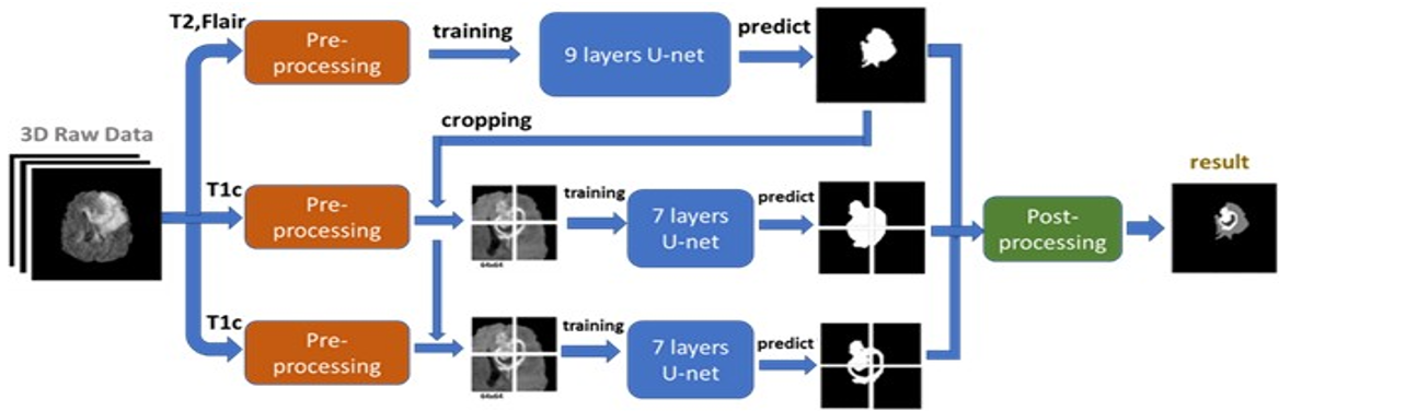
Deep learning techniques have recently demonstrated promising outcomes in the segmentation of brain tumors from MRI images. Due to its capability to handle high-resolution images and segment the entire tumor region, the U-Net model is one of them and is frequently utilized. For the analysis and planning of brain tumors treatments, accurate segmentation of brain tumors using multi-contrast MRI images is essential. Deep learning models including U-Net, PSPNet, DeepLabV3+, and ResNet50 have demonstrated encouraging outcomes in the segmentation of brain tumors. Using the BraTS 2018 dataset, we compare these models in this research. We evaluate the models using a variety of measures, including the Hausdorff Distance (HD), the Absolute Volume Difference (AVD), and the Dice Similarity Coefficient (DSC), and we look into how data augmentation and transfer learning approaches affect the models' performance. The findings demonstrate that the 3D U-Net model performed the best, with a DSC of 0.90, HD of 10.69mm, and AVD of 11.15%. The PSPNet model achieved comparable performance, with a DSC of 0.89, HD of 11.37mm, and AVD of 12.24%. The DeepLabV3+ and ResNet50 models achieved lower performance, with DSCs of 0.85 and 0.83, respectively. Based on the discoveries and analysis, the 3D U-Net model with data augmentation and transfer learning is suggested for brain tumors segmentation utilizing multi-contrast MRI images.
URN:NBN:sciencein.jist.2024.v12.793
Downloads
Downloads
Published
Issue
Section
URN
License
Copyright (c) 2024 Alok Singh Chauhan, Jyoti Singh, Sumit Kumar, Neeru Saxena, Meghna Gupta, Poonam Verma

This work is licensed under a Creative Commons Attribution-NonCommercial-NoDerivatives 4.0 International License.
Rights and Permission
How to Cite


